 Atlas of MRI Brain Anatomy
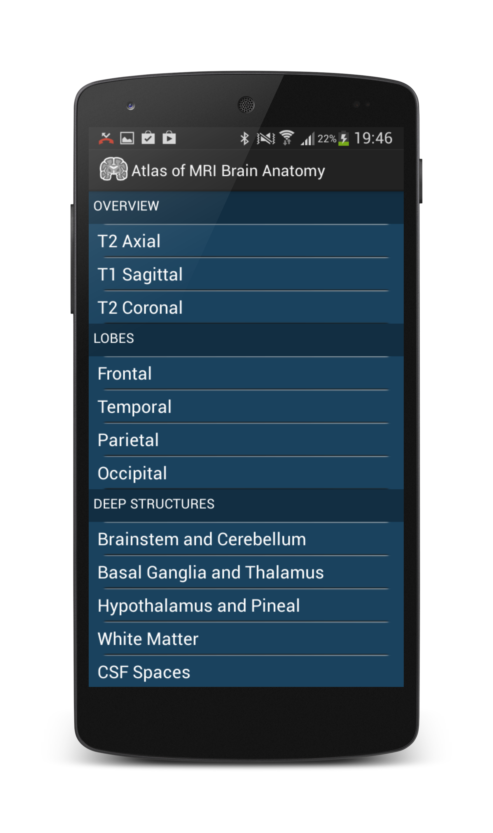
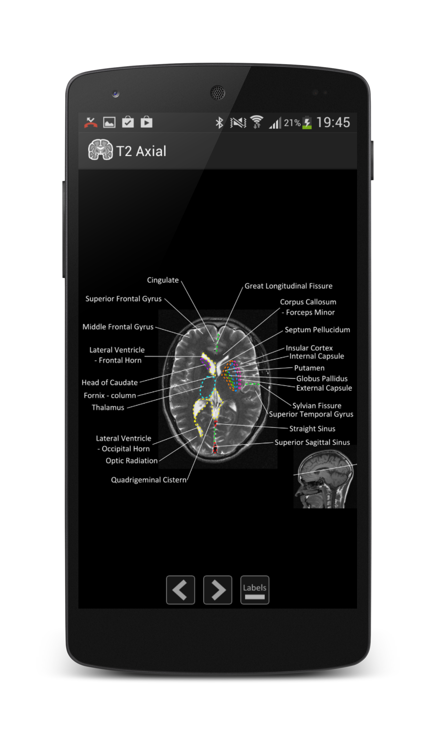
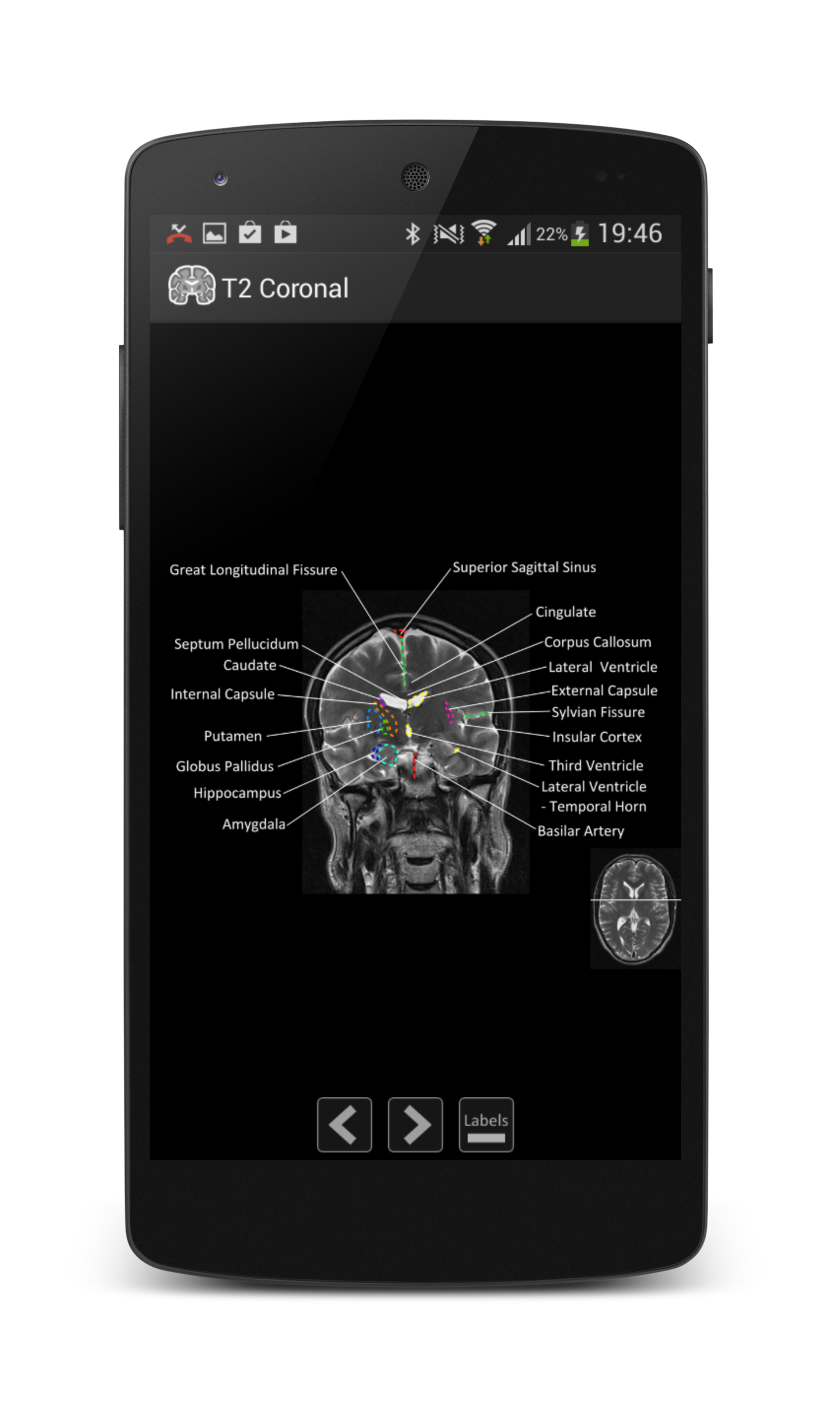

Atlas of MRI Brain Anatomy is the definitive MRI neuroanatomy guide. It contains zoomable cross sectional MRI brain in three planes, labelled in detail covering:- Overview: axial, coronal and sagittal brain sections labelled in moderate detail
- Detailed subsections covering anatomy of individual lobes, brainstems and cerebellum, basal ganglia and thalamus, white matter, CSF spaces and vessels
- Images can be zoomed (double tap or pinch) and labels toggled on or off
- Also includes links to key online and text references.
Atlas of MRI Brain Anatomy is designed for anyone with an interest in neuroanatomy including medical students, neurology trainees and specialists as well as gereneral radiologists or radiological trainees.
Images can be magnified (by pinch or double tap) and labels can be toggled on or off.
Please note that Atlas of MRI Brain Anatomy is intended as an educational anatomy guide only and should not be used for diagnostic purposes. Furthermore, brain anatomy can vary significantly between individuals.